Release date: 2015-11-27
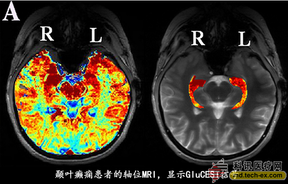
Researchers are experimenting with a powerful imaging tool to locate epilepsy lesions that are difficult to find. Glutamate chemical exchange saturation transfer (GluCEST) is a high-resolution MRI technique that detects how much glutamate is present in the hippocampus. The findings were published in the journal Science Translational Medicine. Glutamate is the most common excitatory neurotransmitter in the central nervous system. Studies have shown that it increases in seizures at onset, during seizures, and at autopsy. "The core hypothesis in epilepsy is that there are some mismatches between how much glutamate inhibition and how much activation there is when someone has a seizure," said Kathryn Adamiak Davis of the University of Pennsylvania.
Davis, the first author of the study, said the new tool is more sensitive than the currently available imaging methods in detecting hippocampus containing epilepsy networks. The study was very small and included only 4 patients with epilepsy, but the researchers said the results were striking.
They are expected to publish this result as they are preparing for a larger study using more sophisticated multi-slice spiral MRI scanning technology than the single-layer technology used in this study. Research coauthors Ravinder Reddy and Hari Hariharan have imaging metabolites and CEST MRI patents using these as biomarkers.
Temporal lobe epilepsy
The authors say that better detection of epileptic lesions can significantly improve the quality of life of patients with care and epilepsy.
Localization-related epilepsy (LRE), also known as partial epileptic seizures, is the most common type of epilepsy that occurs in 80% of drug-resistant patients. In adults, temporal lobe epilepsy (TLE) accounts for 65% of LRE. Medial temporal sclerotherapy can be identified on MRI in 2/3 temporal lobe epilepsy patients and is associated with the most favorable prognosis for epilepsy surgery.
Patients with drug-resistant epilepsy often use multimodal structural and functional imaging for evaluation in surgical planning. In addition to MRI, 18-fluorodeoxyglucose PET-CT and magnetoencephalography are also included. However, these theories do not adequately localize epilepsy lesions in most patients.
About one-third of TLE patients did not detect any lesions in conventional MRI. However, among patients who underwent epilepsy surgery, 87% of patients had histopathological abnormalities, suggesting that the lesion did exist, but current imaging techniques are not sensitive to the detection of lesions.
The new study included 1 male, 3 female patients, with an average age of 40 years, who were identified as non-lesional TLE and included 11 healthy adults as controls (3 males, 8 females, mean age 35 years).
Half of the seizures in patients with epilepsy come from the left hippocampus and the other half from the right hippocampus. All patients were resistant to a variety of anti-epileptic drugs and had tried it, regardless of the use of several drugs.
Each patient underwent GluCEST at the same time point. With the help of CEST technology, researchers can measure glutamate indirectly by measuring the hydrogen concentration in water.
According to the authors, CEST imaging was performed with an MRI scan with a magnetic field strength of 7T. The sensitivity of GluCEST is at least two orders of magnitude higher than traditional MRI for measuring glutamate, and it can also glutamate in vivo at much higher spatial resolution levels than magnetic resonance spectroscopy (MRS) or spectral imaging. Perform imaging.

In this study, researchers can get results by showing a level of the hippocampus, although the techniques they use now can get 60 levels of images. “So we can get an image of the distribution of glutamate concentrations in most brains,†Davis said.
The results showed that in the four patients with epilepsy, the glutamate concentration measured by GluCEST was significantly higher in both the quality and the number in the hippocampus of the epileptic (ipsilateral) compared with the contralateral side. An accurate number can be obtained to create a region of interest, Davis said. "This scan is also seen by blind epilepsy experts who can also identify an increase in glutamate area in all patients."
In the patients studied, the GluCEST signal was significantly different in the ipsilateral (seizure) head from the contralateral side (single tail, P = 0.03). Similar differences in hippocampus, entire hemisphere, and hemispheres that did not include occipital lobe (including major temporal and temporal medial structures) were not statistically significant.
An epilepsy patient was subsequently evaluated for intracranial EEG and right temporal lobe resection. The patient's pathological findings showed that the surgical resection area was consistent with the medial temporal lobe sclerosis. The author said that this "gives GluCEST further confidence." Surgery for the other three patients is under planning.
This promising new approach has several advantages over MRS. MRS is the only technology other than GluCEST that can non-invasively measure glutamate levels in human brain. In addition to higher resolution, it takes less time and is less subject to motion artifacts. GluCEST has a higher spatial resolution than PET-CT, and PET-CT has been used to measure glutamate receptors in healthy controls.
New imaging tools can reduce the need for invasive intracranial monitoring, which results in higher morbidity and mortality and is more expensive. "Now in patients undergoing surgery, we believe that seizures come from the hippocampus, but it is not clear which side, and there is no lesion on MRI. At this time, we often insert intracranial electrodes under the skull and invade. Sexual examination," the author said.
In addition, this check can get information about the prognosis and help doctors develop the best treatment. "Our current MRI misses a small lesion, and this examination of the underlying function of the brain excitability network gives hope to guide us to choose the right treatment, perhaps to free patients from invasive examinations."
Epilepsy affects 65 million people worldwide. About one-third of patients with seizures cannot be controlled by drugs. According to Davis, most antiepileptic drugs work by increasing glutamate inhibition or reducing activation. “Now we believe that all our treatments are adjusting the balance between inhibition and activation.â€
In addition to using a multi-layered approach for a larger study, Davis et al. are expanding their research on all types of drug-resistant epilepsy. They also do some work on animal models using stronger MRI (9.4T).
High level glutamate
Richard Conroy of the project's sponsor, the Institute of Biomedical Imaging and Bioengineering (NIBIB), said that although the scale of the study is small, it still has two important implications.
One is that this study is the first to confirm the previously suspected fact that glutamate levels are elevated in the area of ​​seizures; the second is that this study uses high-intensity imaging to replace MRS-related Some "background features." Conroy added that a typical MRS only shows an area of ​​the brain, which is a "challenge."
Conroy acknowledged that only one patient in the study provided definitive evidence. "Part of the argument is that the level of glutamate found in the scan was abnormally confirmed in the case, but it is only a patient, so it is difficult to be a powerful example."
He also said that another challenge is the 7T MRI scanning device, which is only available in academic centers. Davis said there are currently 20 to 25 such scanning devices across the country.
Conroy also commented that different types of epilepsy classification are not based on lesion location, but on drug resistance. “So the point of the question is, once applied to a larger group of people, will they only see this in the subgroup of the subgroup?â€
Another problem is that he said that MRI with lower magnetic field strength, such as the application of the wider 1.5T and 3T devices, can also detect the difference in glutamic acid. “Once you know what you are looking for, it may become easier to prove in the more common MRI.â€
Source: Medical Pulse
All safety-conscious parents lookout for a solution that can track their child`s school bus location in real-time, and that`s where a School Bus GPS Tracking System comes in.
A real-time bus tracking system provides you the live GPS location of the school bus and tells you whether your child has boarded or de-boarded the bus or not, and at what time.
All this information can be accessed by parents from anywhere, be it their home or workplace.
That`s the whole beauty of a real-time tracking system!
Complete Safety and Security of Kids:
Transportation safety is the primary concern of parents.
The Best school bus tracking system allows school administrators to keep a tab on driver`s speed limits and provides an option to contact your drivers on duty. This gives peace of mind to parents in ensuring the safety and security of their kids.
Monitor assist system,Vehicle tracking system,Vehicle monitoring system,Vehicle remote monitoring system , Vehicle backup camera system,Vehicle driver monitoring system
SHENZHEN SANAN TECHNOLOGY CO.,LTD , https://www.sanan-cctv.com