Character identification
(1) The root bar of the five-plus-pink root is irregular and Zheng or Zheng Zheng barrel-like, some are piece-shaped, length 4-15cm, diameter 0.5-1.5cm, thickness 1-4mm. The outer surface is gray-brown or gray-brown, with irregular cracks or longitudinal wrinkles and long lenticels; the inner surface is white or grayish yellow with fine vertical stripes. Lightweight, crisp, irregular section, gray or grayish yellow. The gas is slightly fragrant and the taste is slightly spicy and bitter.
It is better to have thick skin, aroma, and grayish white cross-section.
(2) Acanthus sinensis is cut into a cylindrical shape or cut into irregular pieces. Thick skin 0.5-1mm. The outer surface is gray-brown and brown, with longitudinal wrinkles, light lenticels, and lateral bulges. Bark dark gray or gray-black; tender stems with thorns, a flat cone, most spalled, punctate or radial oval punctate. Gas is slightly fragrant and tasteless.
(3) The dried root bark is in roll shape, single or double rolls, 7 to 10 cm long, 6 mm in diameter, and 1 to 2 mm thick. The evening surface is gray-brown, with horizontal lenticels and longitudinal wrinkles, and the inner surface is light yellow or yellowish brown. Brittle, easy to break, irregular section; light gray yellow. Gas slightly fragrant, slightly bitter taste. Thick, thick skin, incense, no wooden heart is better.
Microscopic identification
(1) Transverse section of fine root and root bark: 7-14 rows of cork. Cortical cells 2-4 columns scattered with resin. The phloem ray is 1-8 cells wide, the phloem resin channels are arranged in a 3-5(-8) ring layer, the resin channels are oval or oval, the major diameter is 60-225(-420) μm, and the diameter is 14- 24 μm, 4-5 peripheral secretory cells, oblong and rectangular, containing light yellowish secretions, the medial resinous tracts are mostly round and 14-20 μm in diameter. Cortical and phloem parenchyma cells containing calcium oxalate clusters have a diameter of 12-60 μm, coarse corners, few sharp tips, small clusters of ray cells, and constant or dozens of radial alignments. Parenchyma cells contain starch grains, single oval or spheroidal, diameter 8-14μm. The bast fibers in the old root bark have a length of 240-600 μm, and the cells are narrow and the hole Han is obvious.
(2) sessile five plus root bark cross section: cork layer 12-20. Cortical 3-4 columns. The phloem ray has a width of 1-3 rows of cells, a wavy curve, a cell wall with a reticular texture, a plurality of phloem resin pathways, an elliptical or circular shape, a major diameter of 40-116 μm, and 4-8 peripheral secretory cells of a diameter of 32-50 μm. In parenchyma cells, clusters of 8-20 (-40) μm phloem occasionally have single or 2-7 bundled fibers scattered. The fiber length is 220-600μm, diameter is 20-50μm, and the pores are obvious.
Bark cross-section: cork layer thickness of 20-25 columns, some cork cell wall thickening was stratified. Thick-corner cells 5-6 columns. There are fiber bundles in the phloem, scattered between the rays, fiber length 600-1500 (-2000) μm, fewer resin tracts, and more distributed in the outside of fiber bundles, parenchyma cell clusters less than root bark.
Physical and chemical identification
TLC:
(1) Take the product 0.5g, add chloroform 10ml, ultrasonic vibration 30min, take 1ml, for spot use. In addition, β-sitosterol and iso-kaurenoic acid/d-sesamin were used as controls. Each point was on a silica gel G plate, developed with toluene-hexyl alcohol ester-acetone (3.5:0.5:0.5), sprayed with 10% sulfuric acid, and heated with a hot air stream. The test solution was spectrally matched with the reference substance. The spots show the same color spots.
(2) take the product 0.5g, filter paper, add 70% alcohol solution 10ml, soak overnight, ultrasonic vibration in hot water for 30min, the solution is concentrated to dryness, and then add 1ml of methanol to dissolve, for spot use. In addition, take Acanthopanax senticosus D, Clove Berry (or B), Carrot Clam (or A) control, and develop with chloroform-methanol-(6:3:1 lower layer liquid), spray 10% sulfuric acid, and blow hot air. Color development, test solution chromatography in the corresponding position with the control chromatography, should be the same color spots.
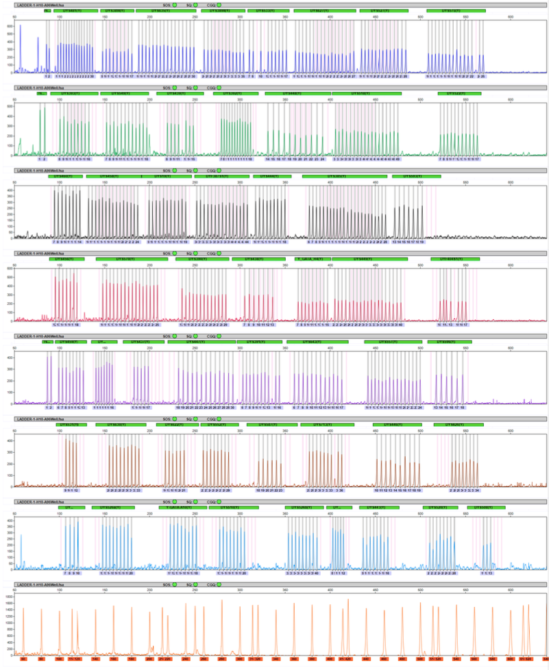
To extract a mixture of DNA fragments, put through a PCR instrument to do a simple purification. Remove the free fluorescent ddNTP single nucleotide, leaving a DNA fragment of a certain length, which can be sequenced on the machine. A polyacrylamide solution is first injected into a hollow capillary during the sequencing process. Then, the polyacrylamide solution was ionized by UV light irradiation to generate a polymerization reaction. The polyacrylamide gel produces a separation effect under the electric field to start the electrophoresis of nucleotide. The electrophoretic movement of short DNA fragments is fast, and the electrophoretic movement of long DNA fragments is slow. The mixture of DNA fragments moves from a negative charge to a positive charge under the action of an electric field in a capillary containing a polyacrylamide gel. The positive end of the capillary is irradiated with a solid-state laser, and a spectroscopic optical sensor records the different fluorescence intensities. Each DNA fragment, when passing through the laser scanning point, has a fluorescent group on it, which will emit a specific fluorescent color.
Because in the previous polymerization reaction process, the starting point of the polymerization reaction starts from a specific primer position. Therefore, the DNA fragment that reaches the laser scanning point of electrophoresis first is the shorter fragment, so its polymerization termination position will be closer to the polymerization start position. Therefore, the fluorescent color reflects which of the bases at its 3' end is A, T, C, and G.
Conversely, the slower the electrophoresis of DNA fragments reaches the laser scanning point, the longer the DNA fragments. As a result, its termination site is farther away from the starting position of the primer. Finally, a map of four colors is obtained.

Fragment Analysis Instrument,Medical Diagnosis Clinical Analyzer,Forensic Testing Dna Sequencer,Capillary Fragment Analysis Genetic Analyzer
Nanjing Superyears Gene Technology Co., Ltd. , https://www.superyearsglobal.com